

Prof. Cordula Poulsen Nautrup Ultrasound Laboratory for Students
Practical training of sonography and echocardiography under supervision of instructors or for self study for veterinary students and postgraduate veterinarians during their doctoral thesis
Since 2007 a modern ultrasound laboratory for students is well-established at the Chair of Anatomy, Histology and Embryology of LMU’s Faculty of Veterinary medicine. The laboratory was founded and opened with financial support from student fees and is now financed by grants of the Bavarian Government for universities and supported by industry. In this Lab students and postgraduates can learn and practice sonography and echocardiography practically.
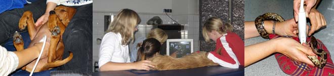
The ultrasound laboratory is equipped with four modern middle- to high class ultrasound systems. Almost all sorts of ultrasound probes and frequencies are available, so ultrasound examination of almost every organ – including the heart - of dogs, cats, sheep, goat, piglet, calf, rabbits, mice and reptiles is possible. Sonography of anatomic specimen of limbs of cows and horses is possible as well. The ultrasound systems are capable of high-definition one- and two-dimensional imaging, as well as all common color flow Doppler and pulsed- and continuous wave Doppler imaging.
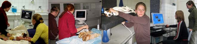
Three specially-skilled veterinarians – with long-term experience in ultrasound and ultrasound simulation – supervise the students in handling the ultrasound devices and teach them how to carry out ultrasound examinations properly. Computer work stations with multimedia ultrasound teaching software and several books on ultrasound examinations are as well available.
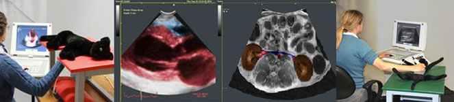
Since 2015 the world’s first veterinary ultrasound simulations system can additionally be used by the students. With this simulation system an almost real ultrasound examination of “healthy” and “sick” cat models can be carried out. It is as well possible to compare ultrasound images and cross-sectional views with the help of a split screen view. Additionally the organs and structures can be labeled with their correct name for teaching purposes when required. So especially for beginners, it is easier to find and identify the correct anatomic structures. Auscultation of the cat’s heart can as well be practiced with the help of this simulation system.
Students who want to learn and practice ultrasound at the Professor Cordula Poulsen Nautrup ultrasound lab should:
- mistudy at least in the 5th semester
- come together in small groups of 2-4
- have an animal that wants to be examined with ultrasound (only small animals, no horses, cows or other large animals because of the dimensions of the laboratory)
- No animal is sedated, most of them get used to ultrasound examination until the second visit
- have a two-hour time slot
- make an appointment with the instructors
- for ultrasound simulation courses, come together in small groups of 2-4 as well and make an appointment
Appointments:
- Directly contact the instructors/lecturers
Dr. Stefanie Weber
Dr. Inga Wölfel
Dr. Elisabeth Zandt - by phone: 089-2180-2568
- by email: ultraschall.studvet@anat.vetmed.uni-muenchen.de